The question
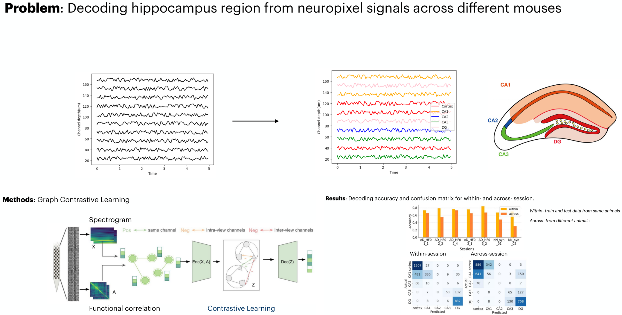
In multi-day recordings and in deep brain stimulation, you need to know exactly where each electrode sits, often down to a thin layer of the hippocampus. Each current approach misses something. Atlas-guided insertion is thrown off by differences between animals. CT and MRI can’t resolve small subregions. Histology only works after the experiment ends.
The hippocampus has a distinctive layered structure, and each layer has its own electrical behavior. Can we work out each channel’s location “blind,” from the recordings alone?
What we did
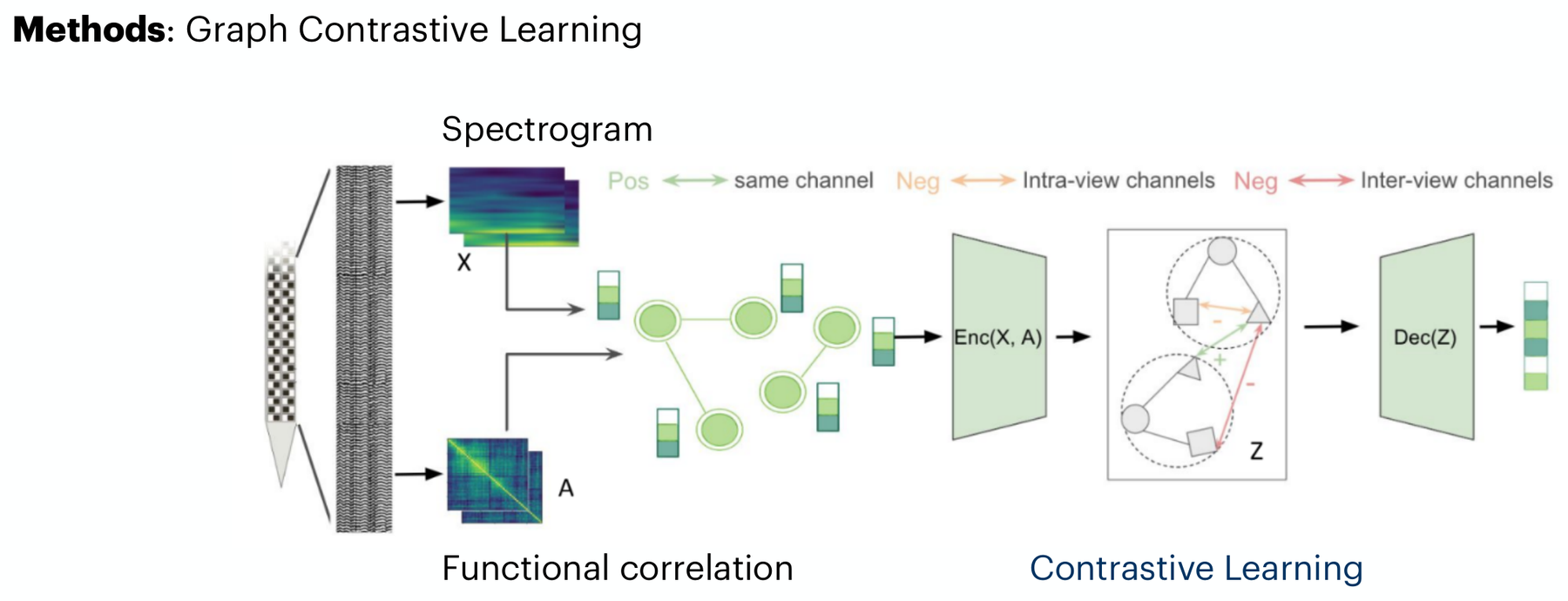
We used high-density Neuronexus probes with 1024 channels over 8 shanks, recorded in 7 mice. For every pair of channels we computed how strongly their local field potentials (LFPs) correlate. That gives each channel a profile of how it relates to all the others. We combined these correlation patterns with spectrograms of each channel.
Recordings from different animals and sessions don’t line up directly. We aligned them into a shared space in two ways: a simple rotation and shift per session, and a neural network trained with supervised contrastive learning. A probabilistic decoder then predicts each channel’s region (cortex, CA1, CA2, CA3, or dentate gyrus).
What we found
1. Brains from different animals share the same shape
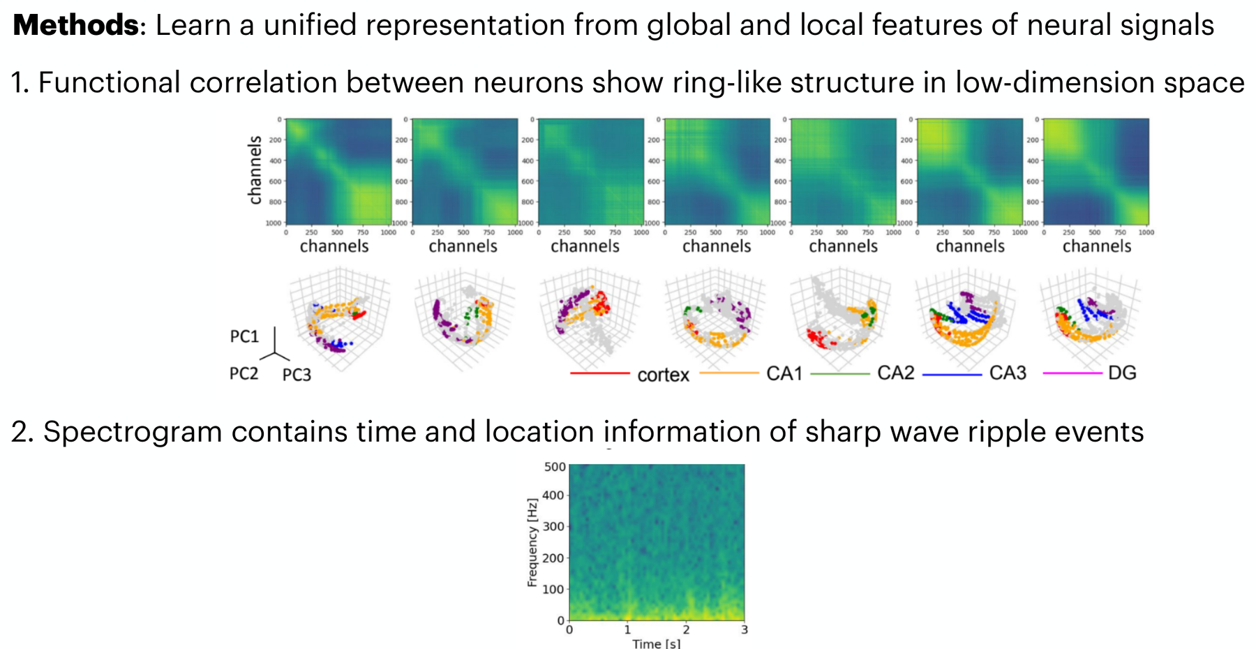
In a low-dimensional space, the correlation patterns form a ring-like structure, with channels ordered by region. That shape is shared across sessions and animals, apart from simple shifts and rotations. This is what makes alignment, and cross-animal decoding, possible.
2. Accurate within an animal, and still useful across animals
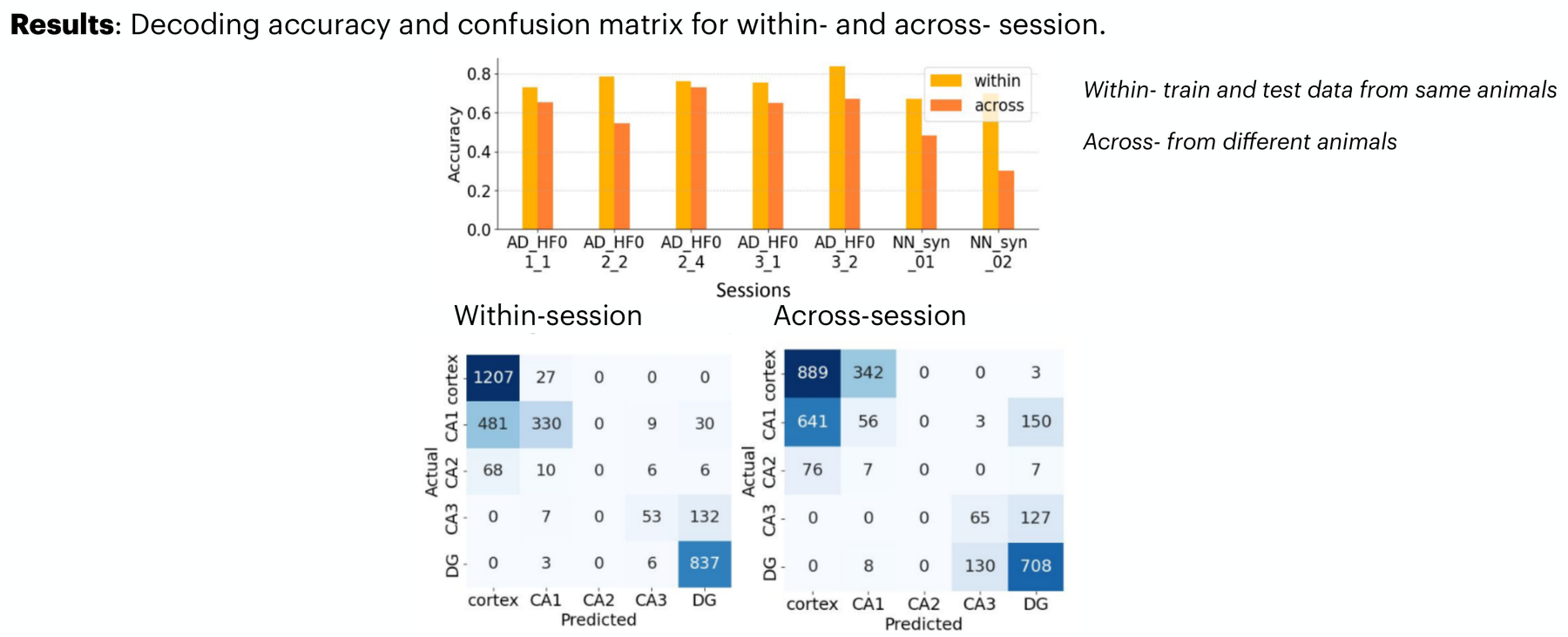
Within a session, the model labeled all 1024 channels with 93% accuracy using only 5% labeled channels, outperforming a baseline adapted from LOLCAT. Across sessions, with minimal labels in the new session, it reached 71%. Its predictions reproduced the hippocampus’s layered structure even though the model was never told about it.
This project led to Lfp2vec, a self-supervised model that scales the idea up to raw LFP across labs and species.
My take
Links
BibTeX
@misc{he2024blind,
  title        = {Blind In Vivo Localization of Microelectrode Arrays via Functional
                  Correlation Patterns in the Mouse Hippocampus},
  author       = {He, Tianxiao and Maslarova, Anna and V{\"o}r{\"o}slakos, Mih{\'a}ly and
                  Li, Chenyi and Patel, Malhar and Hung, Allen and Ramanathan, Nalini and
                  Liu, Yurong and Afrasiyabi, Arman and Hurwitz, Cole and
                  Buzs{\'a}ki, Gy{\"o}rgy and Varol, Erdem},
  howpublished = {Poster, Society for Neuroscience Annual Meeting (SfN), Chicago, IL},
  year         = {2024}
}